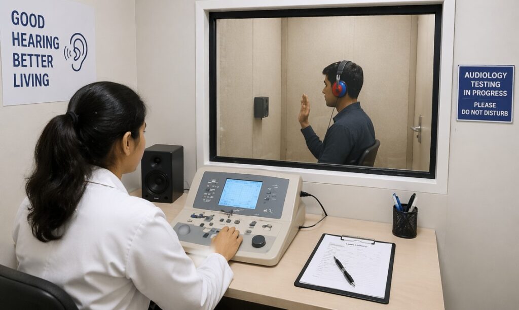
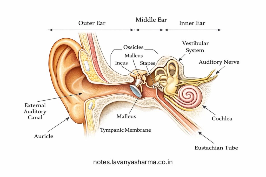
The human ear is an important sense organ, and we have two ears located on either side of our face. The part of the ear that we can see from outside is only a small portion of the entire ear structure. The ear, also known as the auditory system, is divided into three main parts: the outer (external) ear, the middle ear, and the inner ear. Each part has a specific role in the process of hearing. The inner ear is connected to the auditory nerve, which carries sound signals to the brain through the auditory nervous system.
Anatomy
External Ear
The outer or external ear is the visible part of the ear and consists of the pinna (auricle) and the ear canal. The pinna is a flap-like structure attached to the side of the head at an angle of about 30 degrees. It acts like a funnel, collecting sound waves and directing them into the ear canal (external auditory meatus) towards the tympanic membrane (eardrum). The word “pinna” comes from Latin, meaning “wing.”
The surface of the pinna is uneven and contains several structures such as pits, grooves, and depressions. The deepest depression is called the concha, which helps in directing sound into the ear canal. The outer rim is called the helix, and a parallel inner ridge is known as the antihelix. The space between them is called the scaphoid fossa. Near the ear canal is a small flap called the tragus, and opposite to it is the antitragus, separated by the intertragal notch. The lower soft part of the ear is called the lobule or earlobe.
The ear canal (external auditory meatus) is a curved tube about 25 mm long and 8 mm wide. It carries sound waves from the pinna to the eardrum. The canal is partly cartilaginous (outer part) and partly bony (inner part). The cartilaginous portion can change shape with jaw movement, while the bony part is fixed. At birth, the bony part is not fully developed and completes its development by around 3 years of age.
Middle Ear
The external auditory meatus leads to the tympanic membrane or eardrum, which marks the beginning of the middle ear. The middle ear, also called the “tympanum,” is an air-filled cavity that resembles a drum. It consists of the tympanic membrane, the middle ear cavity, and structures such as ossicles, muscles, and a mucous membrane lining.
The tympanic membrane is a very thin (about 0.1 mm), cone-shaped structure placed obliquely and slightly pushed inward at its center. It is fully developed during fetal life and forms the lateral wall of the middle ear. The middle ear cavity has six walls: lateral, medial, anterior, posterior, roof, and floor.
Inside the middle ear are three tiny bones called ossicles—malleus, incus, and stapes—which help in transmitting sound vibrations from the eardrum to the cochlea in the inner ear. The Eustachian tube connects the middle ear to the nasopharynx and plays an important role in maintaining equal air pressure on both sides of the eardrum and draining secretions from the middle ear.
When the air pressure on both sides of the eardrum is equal, sound is transmitted efficiently. However, during situations like high altitude, pressure imbalance can occur, making the eardrum stiff and causing a blocked ear sensation. The Eustachian tube helps to equalize pressure during activities like yawning, chewing, and swallowing. If it does not function properly, it may lead to fluid buildup and infections in the middle ear.
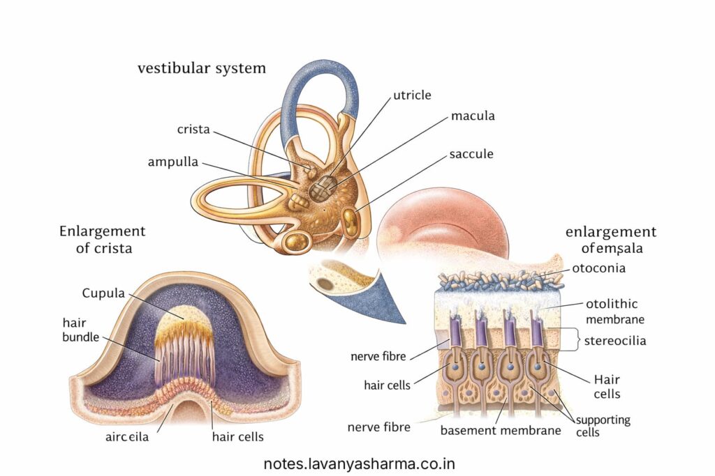
Inner Ear and Vestibular System
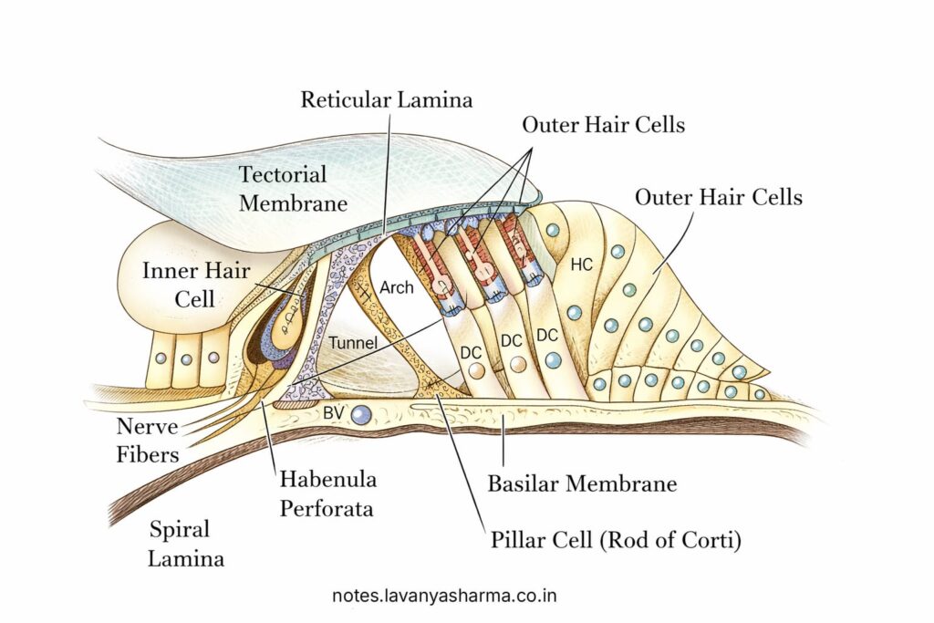
The inner ear is located within the temporal bone of the skull and consists of a system of fluid-filled tubes. It includes two main structures: the bony labyrinth and the membranous labyrinth. The bony labyrinth is filled with a fluid called perilymph, while the membranous labyrinth inside it contains endolymph. The membranous labyrinth has delicate structures that include the organ of Corti, which contains specialized sensory cells called hair cells responsible for hearing.
The inner ear has three main parts. The cochlea, which is spiral or snail-shaped, is the organ of hearing. The semicircular canals help in maintaining balance, and the vestibule connects the cochlea and semicircular canals. The vestibule contains structures called the utricle and saccule, which also play a role in balance.
The inner ear is connected to the middle ear through two openings called the oval window and the round window. The oval window is located just behind the stapes (a middle ear bone). Inside the cochlea, there are two types of hair cells—inner hair cells and outer hair cells—which are located on the basilar membrane and help in detecting sound.
These hair cells are connected to the auditory nerve (8th cranial nerve), which carries sound signals to the brain. The signals pass through important centers such as the cochlear nucleus, superior olivary complex, and inferior colliculus. Finally, the sound is processed and understood in the auditory cortex, which is located in the temporal lobe of the brain.
Auditory Nervous System
The auditory nervous system is responsible for carrying sound information from the ear to the brain for understanding. The auditory nerve, also known as the 8th cranial nerve, plays a key role in this process. The cell bodies of this nerve are located in the spiral ganglion, and its dendrites are connected to the sensory hair cells in the organ of Corti.
The axons of the auditory nerve carry sound signals to the brainstem and enter at the level of the pons, where they first connect with the cochlear nucleus. From there, the sound information travels through several important centers, including the superior olivary complex, lateral lemniscus, inferior colliculus, and medial geniculate body.
Finally, the auditory signals reach the auditory cortex in the brain, where the sound is analyzed for its meaning and content.